Schematic Diagram Of Blood Circulation In Human Body
Part 3 (Blood Platelet) — Blood platelets are the initiators of blood clotting. (d) The average life span of a red blood cell (RBC) is about 120 days. (e) Fibrinogen. Question 2. Given below is a highly schematic diagram of the human blood circulatory system.

May 3, 2015 February 23, 2021 gccexchange 0 Comment
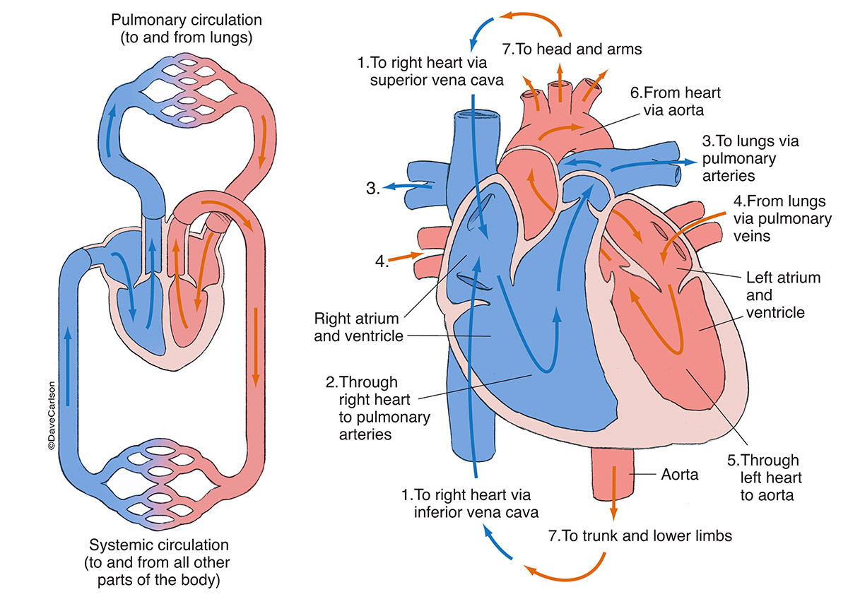
Process of blood circulation: Deoxygenated blood from the body cells are carried by the veins to the heart. The right auricle receives the blood and pumps it (through the tricuspid valves) into the right ventricle. From here, pulmonary artery transports it to the lungs where it gets oxygenated. From the lungs the oxygenated blood is carried by.

Heart Anatomy BIO103 Human Biology
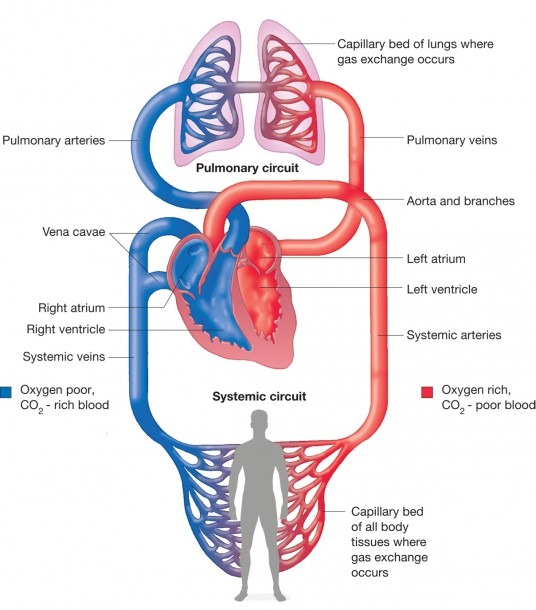
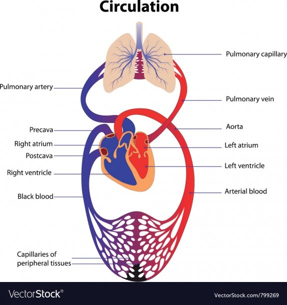
systemic circulation: The part of blood circulation that carries oxygenated blood away from the heart, to the body,. Alveoli: A diagram of the alveoli, showing the capillary beds where gas exchange with the blood occurs. Pulmonary circuit: Diagram of pulmonary circulation. Oxygen-rich blood is shown in red; oxygen-depleted blood in blue.

How Chiropractic Care Has Been Shown to Improve Blood Circulation
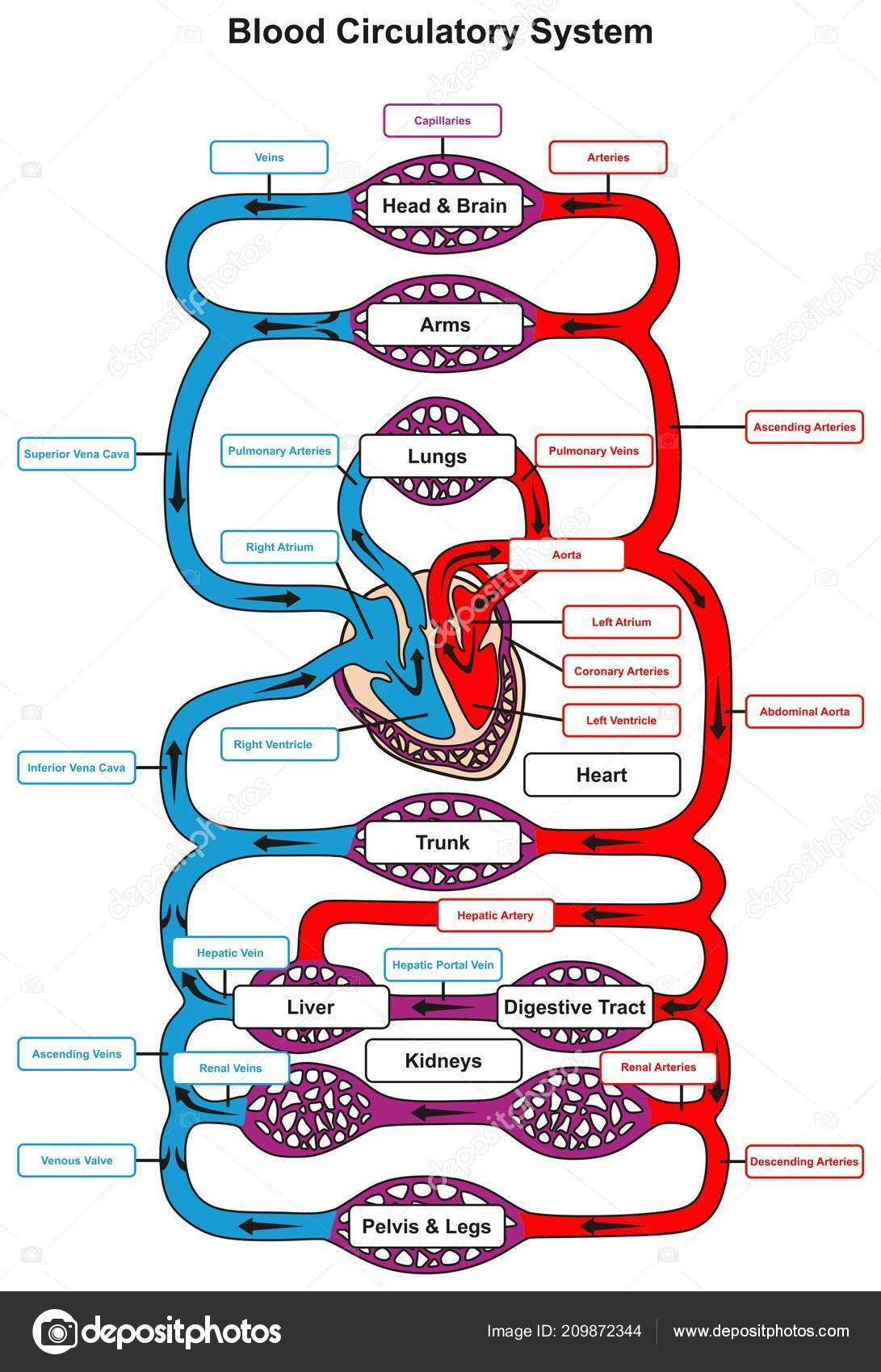
Blood Circulatory System: Types, Diagram, Working, Open vs Closed Circulation, Structure and Functions. Circulatory Systems are bodily systems used to transport nutrients, blood, gases, wastes and other materials of the body to and fro from one part to another part. Circulatory systems are present in the simplest organisms like sea sponges.

Explain the circulation of blood in human body with the help of a schematic diagram
Diagram of the Human Circulatory System (Infographic) Infographics. By Ross Toro. published 29 August 2013. Find out all about the blood, lungs and blood vessels that make up the circulatory system.
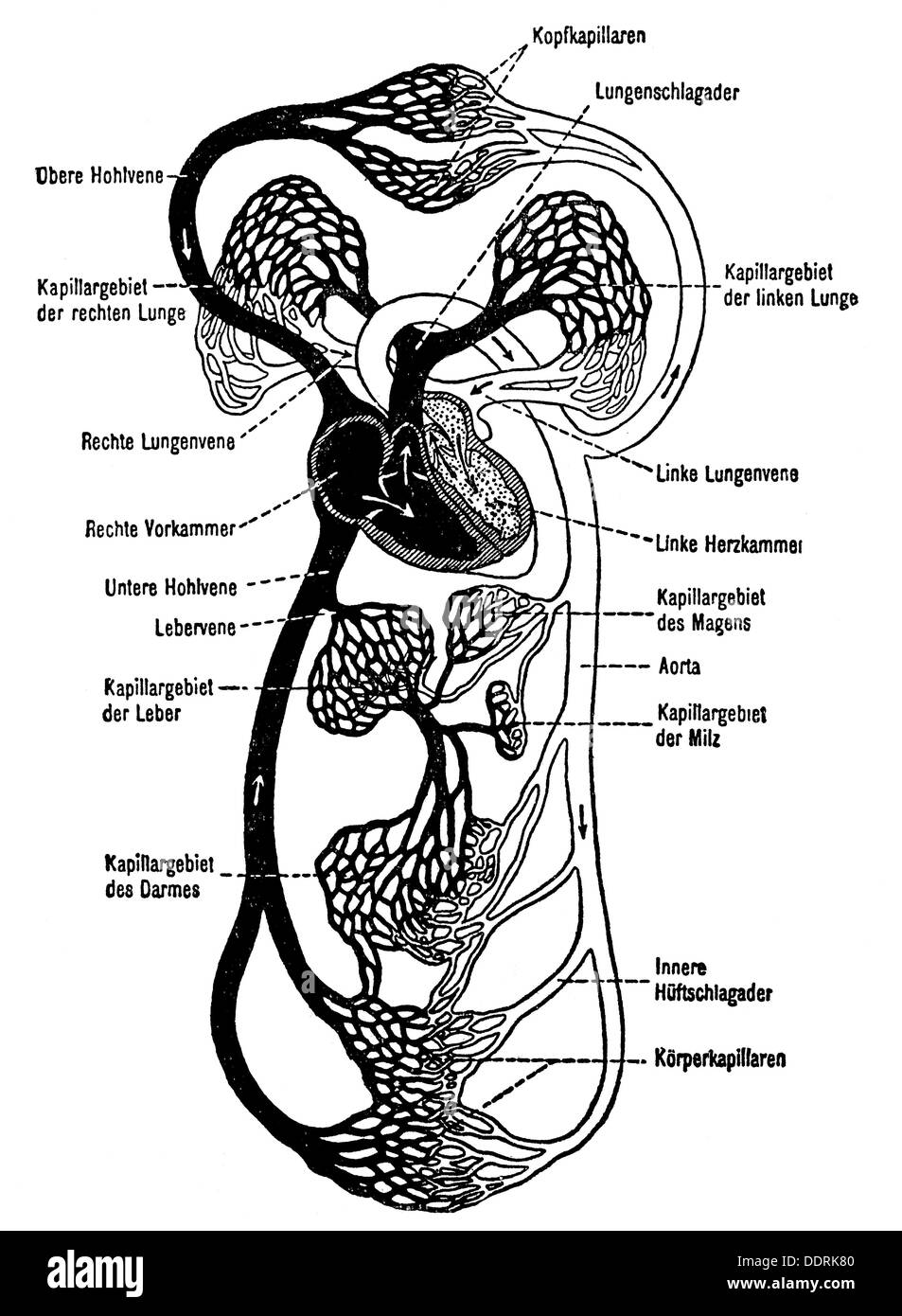
medicine anatomy blood circulation schematic diagram drawing 20th Stock Photo 60141648 Alamy
Blood then enters the left ventricle to be circulated again. Pulmonary circuit: Diagram of pulmonary circulation. Oxygen-rich blood is shown in red; oxygen-depleted blood in blue. Distribution of blood can be modulated by many factors, including increasing or decreasing heart rate and dilation or constriction of blood vessels.

Biology Blood circulation
This Osmosis High-Yield Note provides an overview of Renal Blood Flow Regulation essentials. All Osmosis Notes are clearly laid-out and contain striking images, tables, and diagrams to help visual learners understand complex topics quickly and efficiently. Find more information about Renal Blood Flow Regulation: Regulation of renal blood flow.

3 A schematic overview of the cardiovascular system with the heart as... Download Scientific
These comprise arteries, veins, and capillaries. The primary function of blood vessels is to transport oxygenated blood and nutrients to all parts of the body. It is also tasked with collecting metabolic wastes to be expelled from the body. Most circulatory system diagrams do not visually represent its sheer length.

1 A schematic diagram of the blood circulation system in the human body... Download Scientific
In double circulation, there are two pathways in which blood flows. They are: 1) Pulmonary pathway 2) Systemic pathway. The pulmonary circulation or pathway carries the deoxygenated blood from the right side of the heart to the lungs. Exchange of oxygen and carbon dioxide takes place in the lungs and the blood is now oxygenated (with oxygen).
Blood Circulatory System Human Body Infographic Diagram Heart Pumping All ⬇ Vector Image by
Download scientific diagram | Schematic diagram of the blood circulatory system. There are three major types of blood vessels: arteries, veins and capillaries. Capillaries are consisted of a layer.
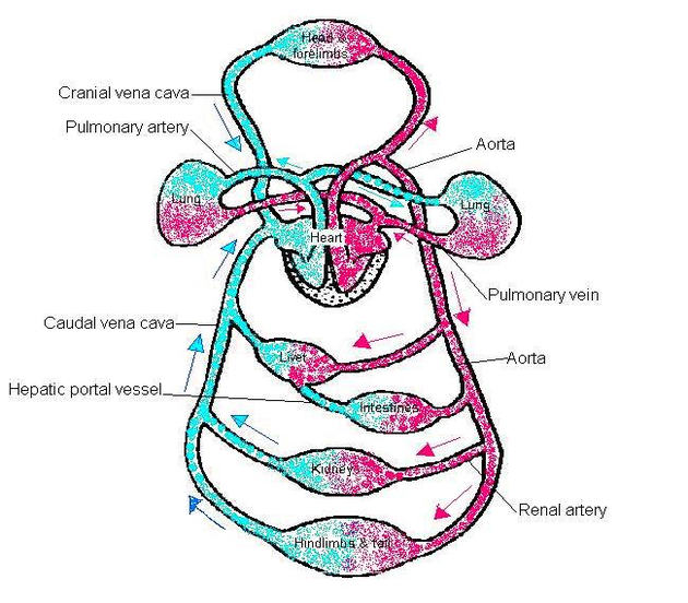
Diagram blood circulation labeled Anatomy System Human Body Anatomy diagram and chart images
Step 1 and 6 involve a blood vessel, which makes sense as this is how blood enters and exits that side of the heart. Steps 2-5 involve a chamber, valve, chamber, and valve. So if you remember this general pattern, it will help you recall the order in which blood flows through each side of the heart.

Double Circulation Blood Circulation in Humans BYJU'S
15.3A: Anatomy of Human Circulatory System. The circulatory system is an organ system that permits blood to circulate and transport nutrients (such as amino acids and electrolytes), oxygen, carbon dioxide, hormones, and blood cells to and from the cells in the body to provide nourishment and help in fighting diseases, stabilize temperature and.
Cardiovascular Networks Generalized Carlson Stock Art
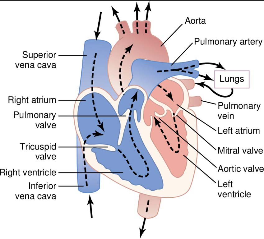
Blood enters the heart through two large veins, the inferior and superior vena cava, emptying oxygen-poor blood from the body into the right atrium of the heart. As the atrium contracts, blood flows from your right atrium into your right ventricle through the open tricuspid valve. When the ventricle is full, the tricuspid valve shuts.
The Blood Circulation in Heart Ishwaranand
This lesson includes the main parts of the heart anatomy and a diagram of the heart and blood flow. Updated: 11/21/2023 Table of Contents. Blood Flow. As we mentioned, blood follows a one-way.
Schematic Diagram Of Blood Circulation In Human Body
Figure 1 - Schematic diagram of the brain blood circulation: 1, Aortic Arch; 2, brachiocephalic artery; 3, common carotid artery; 4, posterior inferior cerebellar.

Human Circulatory System GCSE Biology Revision Notes
The circulatory system works thanks to constant pressure from the heart and valves throughout the body. This pressure ensures that veins carry blood to the heart and arteries transport it away.