
Chilomastix mesnili Medical Laboratories
Chilomastix mesnili is a non-pathogenic [1] member of primate gastrointestinal microflora, commonly associated with but not causing parasitic infections. It is found in about 3.5% of the population in the United States. In addition to humans, Chilomastix is found in chimpanzees, orangutans, monkeys, and pigs. It lives in the cecum and colon.

Chilomastix mesnili Parasitology world
Infection by Chilomastix mesnili was determined by PCR method. Results: We identified nonpathogenic bacteria such as Proteus mirabilis and Escherichia coli in feces of normal common marmosets..

CHILOMASTIX MESNILI PDF
Chilomastix mesnili is a nonpathogenic flagellate that is often described as a commensal organism in the human gastrointestinal tract. Life Cycle View Larger The cyst stage is resistant to environmental pressures and is responsible for transmission of Chilomastix. Both cysts and trophozoites can be found in the feces (diagnostic stages) .

Chilomastix mesnili a photo on Flickriver
Molecular diagnosis Extraction of Parasite DNA from Fecal Specimens Morphologic comparison of intestinal parasites Serum/Plasma Specimens Safety Specimen Requirements Specimen Submission Detection of Antibodies Antibody Detection Test Other Specimens Shipment Tissue Tissue specimens for free-living amebae (FLA) Isolation of Leishmania organisms
Chilomastix sp. Protozoa MONSTER HUNTER'S GUIDE TO VETERINARY
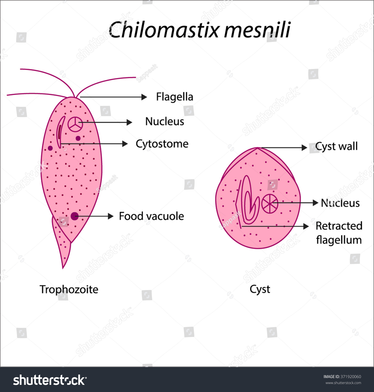
Chilomastix mesnili. Trophozoite and cyst in fecal smear (trichrome stain, oil immersion). A larger trophozoite and a smaller cyst are seen side by side. The trophozoite has the typical pyriform shape and a nucleus with a large, central karyosome. The cyst is more rounded than lemon-shaped and the karyosome is seen in the nucleus. Trophozoites.

10 Practical Parasitology Chilomastix Mesnili Cyst Stage YouTube
Chilomastix mesnili is of cosmopolitan distribution although found more frequently in warm climates. It is thought to be non-pathogenic although the trophozoite has been associated with diarrhoeic stool. Morphology of the cyst The cyst is 6-9 m It has a large single nucleus with a large karyosome.

Chilomastix mesnili YouTube
Infection by Chilomastix mesnili was determined by PCR method. Results: We identified nonpathogenic bacteria such as Proteus mirabilis and Escherichia coli in feces of normal common marmosets. Interestingly, C. mesnili was isolated from a healthy common marmoset by fecal centrifugation concentration and PCR.
Chilomastix mesnil,cyst and trophozoite Royalty Free Stock Vector
Chilomastix exists as a cyst stage that is responsible for transmission and a trophozoite stage which is also known as the feeding stage. Transmission occurs via the fecal-oral route when water contaminated with feces that contain Chilomastix cysts is ingested. [4]

PPT 2013 PARASITOLOGY Lynnegarcia2verizon PowerPoint
Chilomastix cuniculi is a non-pathogenic organism observed in the cecum of the rabbit. The trophozoite is pyriform with three anterior flagella, a large cytosomal groove near the anterior end and an anterior nucleus. The trophozoite ranges in length from 3-20 µm ( Pakes and Gerrity, 1994 ).
Chilomastix sp. Protozoa MONSTER HUNTER'S GUIDE TO VETERINARY
representatives of the genus Chilomastix. trophozoites with single nucleus; 3 anterior flagella and a fourth small flagellum within the cytostomal groove trophozoites with oblique, spiral groove lemon-shaped cysts; mononucleate cytostomal fiber prominent and hook-like in both trophozoite and especially cyst (Shepherd's crook) representative species
Chilomastix mesnili Trophozoites YouTube
Abstract. Scanning electron microscopy of Chilomastix mesnili shows that the cysts are lemon-shaped with one end broadly rounded and the other conical. The trophozoite has five flagella coming out of the anterior end. Four of these are free and the fifth is attached to the body by an undulating membrane. The undulating membrane extends along.

Chilomastix mesnili Medical Laboratories
This photomicrograph of an iodine-stained specimen, revealed some of the ultrastructural morphology exhibited by a flagellated, Chilomastix mesnili trophozoite. Note the organism's distinctly visible cytostome. Trophozoites are pear-shaped and usually measure 6-24 µm in length.

Trofozoitos de Chilomastix mesnili YouTube
Regarding its phylogeny, Chilomastix mesnili belongs to the family Tetramitid~e, the ord Polymastigina, r and the class Mastigophora. The habitat of the parasite is th sm~ll intestine ofman. It is often the cause ofchronic orintermittent diarrhea. Cases ofinfec-tion by Chilomastix in man have been reported from nearly every locality in the world.

pyriform trophozoite
The trophozoites of C. mesnili are also pear-shaped and measure from 6 to 24 µm in length and 4 to 8 µm wide. The single nucleus usually has a prominent karyosome. The anterior flagella are difficult to see. The oral groove (cytostome) is sometimes seen near the nucleus. The image on the left is an iron hematoxylin stain (1000x). Wet Mounts

Pin on Parasitology
This paper reports a case with Chilomastix mesnili infections, and summarizes the diagnosis and treatment with traditional Chinese medicine.. Traditional Chinese Medicine; Trophozoite. Publication types Case Reports MeSH terms Drugs, Chinese Herbal* Humans Medicine, Chinese Traditional Protozoan Infections*.
Chilomastix sp. Protozoa MONSTER HUNTER'S GUIDE TO VETERINARY
The undulating membrane of Trichomonas and the spiral groove of Chilomastix may not be visible in all cases. Cryptosporidium oocysts can be demonstrated in acid-fast stains. Table 3: Differential Morphology of Protozoa Found in Stool Specimens of Humans: Amoebae-Trophozoites