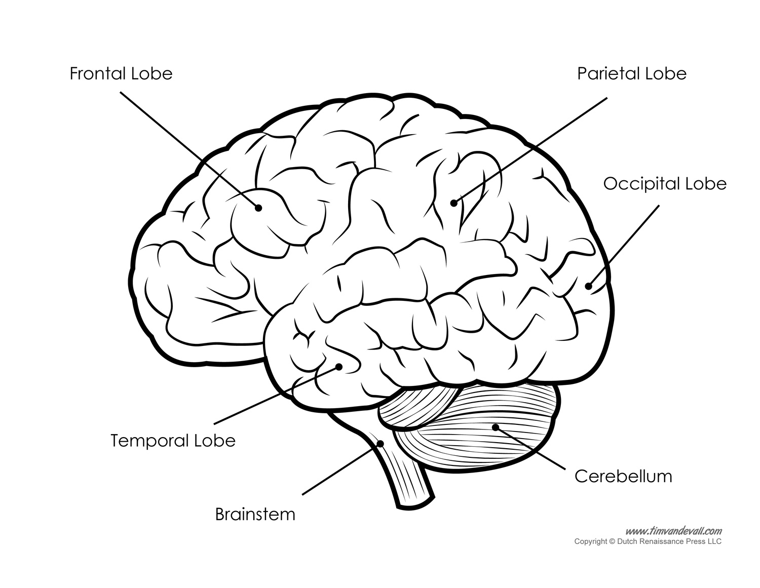
Brain Diagram Labeled BW Tim's Printables
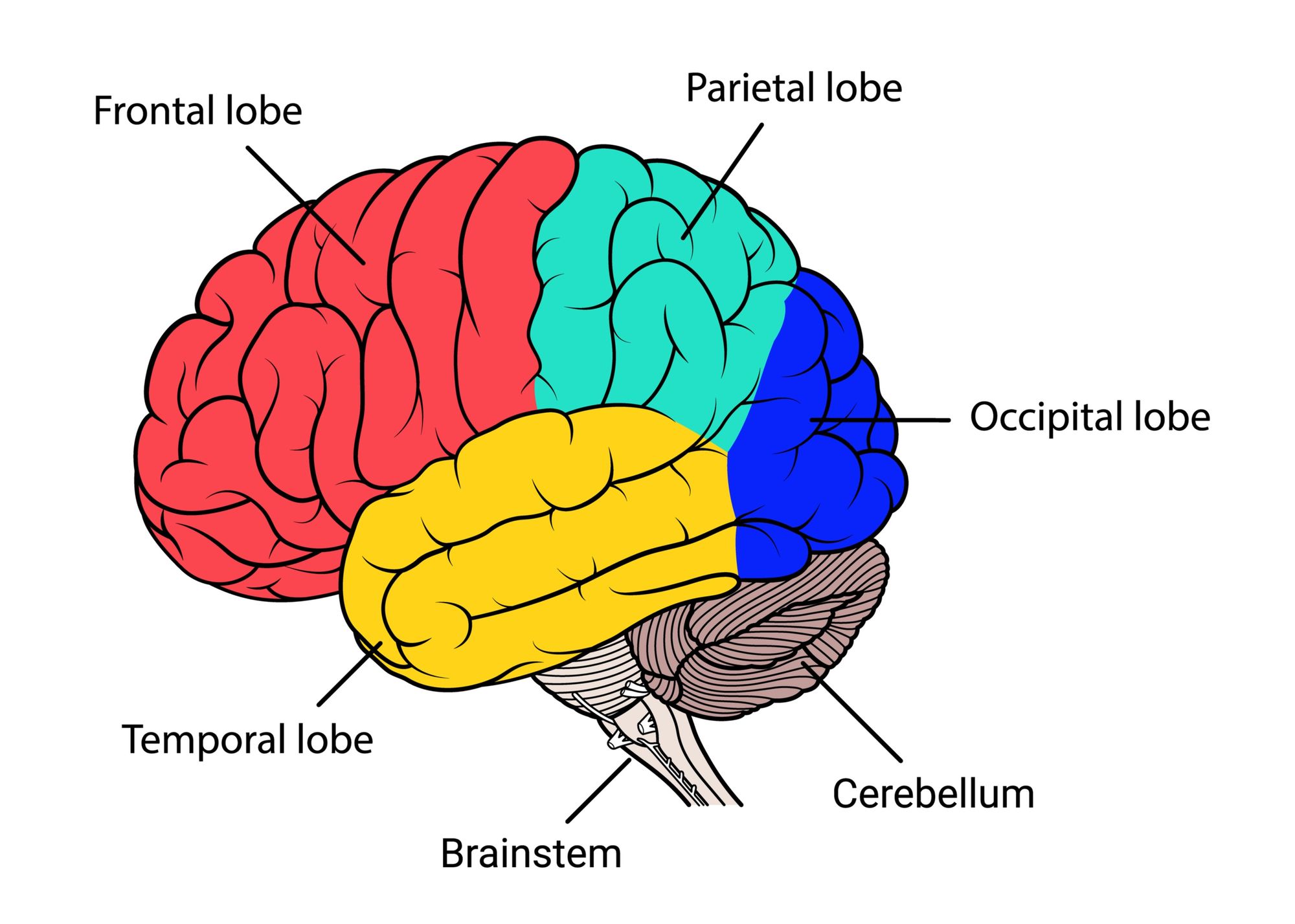
The labeled human brain diagram contains labels for: The frontal lobe, parietal lobe, temporal lobe, occipital lobe, cerebellum, and brainstem. The diagram is available in 3 versions. The first version is color coded by section. The second version is the natural color of the human brain, and the third version is black and white.

Image result for labeled diagram of the brain Brain diagram, Human brain, Brain pictures
It contains 8% proteins 1% carbohydrates, 2% soluble organics and 1% insoluble salts. More than half of the neurons in the brain are found in the cerebellum and only 10% neurons make up the brain. 85% of the brain is cerebral cortex, divided as, 41% frontal lobe, 22% temporal lobe, 19% parietal lobe and 18% occipital lobe.
12 Best Images of Human Brain Diagram Worksheet Human Brain Anatomy Coloring Page, Brain
Brain Basics: Know Your Brain The brain is the most complex part of the human body. This three-pound organ is the seat of intelligence, interpreter of the senses, initiator of body movement, and controller of behavior. Lying in its bony shell and washed by protective fluid, the brain is the source of all the qualities that define our humanity.
Brain Jack Image Brain Diagram
What is a neuron? Nervous system Central nervous system Cerebrum and cerebral cortex Subcortical structures Brainstem Cerebellum Spinal cord Meninges Ventricles and CSF Brain blood supply Peripheral nervous system Cranial nerves Spinal nerves Neural pathways and spinal cord tracts Ascending pathways Descending pathways Sources Related articles

detailed brain diagram awesome detailed brain anatomy Brain diagram, Brain anatomy, Human
MedicalRF.com/Getty Images The cerebral cortex is the part of the brain that makes human beings unique. Functions that originate in the cerebral cortex include: Consciousness Higher-order thinking Imagination Information processing Language Memory Perception Reasoning Sensation Voluntary physical action
PostStroke Dizziness How Vestibular Therapy Can Help
Neurons Glial Cells Cranial Nerves The brain receives information from sensory receptors and sends messages to muscles and glands. It is the center of all conscious awareness and is divided into different lobes with different functions. It contains the cerebrum, about 85% of the total mass.
Free Brain Diagram, Download Free Brain Diagram png images, Free ClipArts on Clipart Library
This brain labeling activity was created for remote learners as an alternative to the labeling and coloring worksheet we would traditionally do in class. Instead of coloring and labeling on printouts, students use google slides to drag labels to the images or type the answers into text boxes. The slides do not have labeled diagrams but does.

Brain Images Labeled
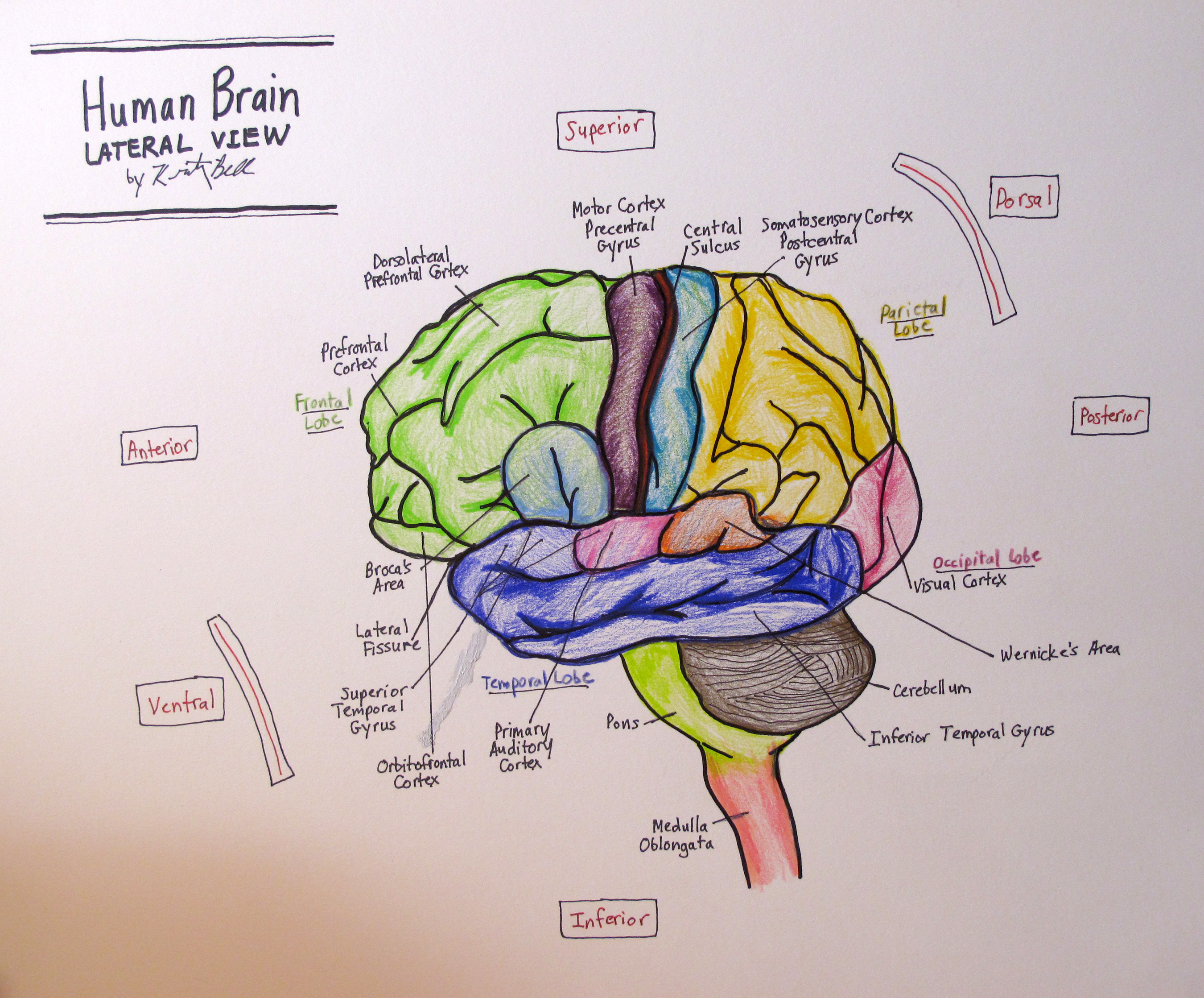
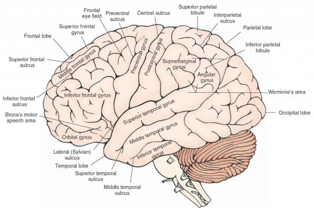
[Lateral views of the brain - labeled diagram]Looking at the brain from the lateral view we can see the frontal, temporal, parietal and occipital lobes. There are several important gyri and sulci that are visible from these two perspectives. The central sulcus separates the frontal from the parietal lobe (and the precentral gyrus from the.
Label The Brain Anatomy Diagram Diagram For You
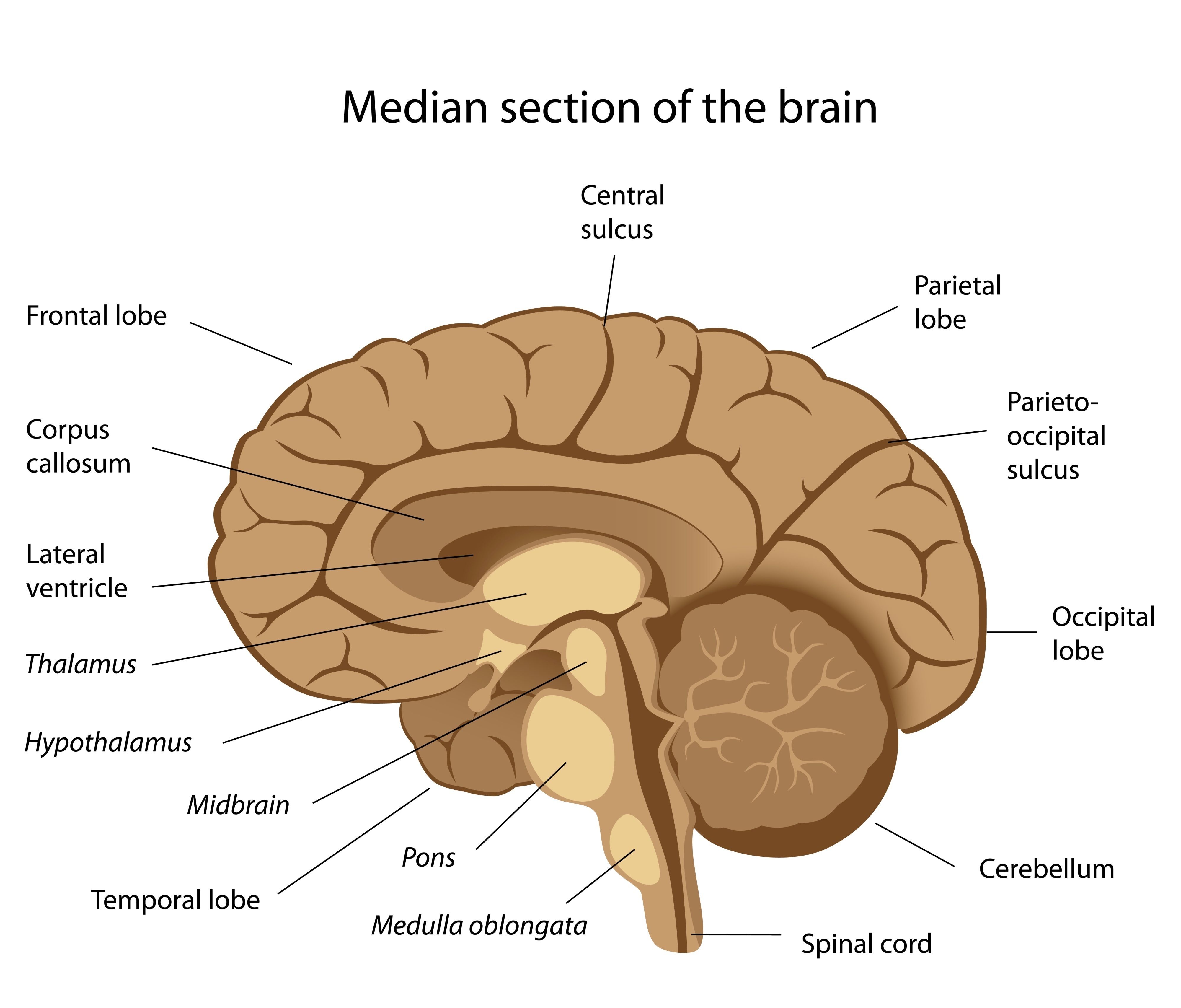
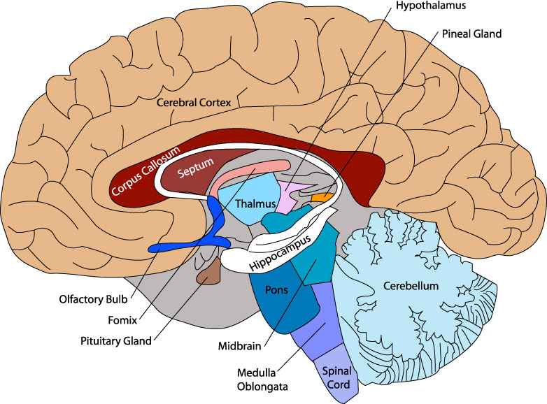
The Brain. Read the definitions below, then label the brain anatomy diagram. Cerebellum - the part of the brain below the back of the cerebrum. It regulates balance, posture, movement, and muscle coordination. Corpus Callosum - a large bundle of nerve fibers that connect the left and right cerebral hemispheres.
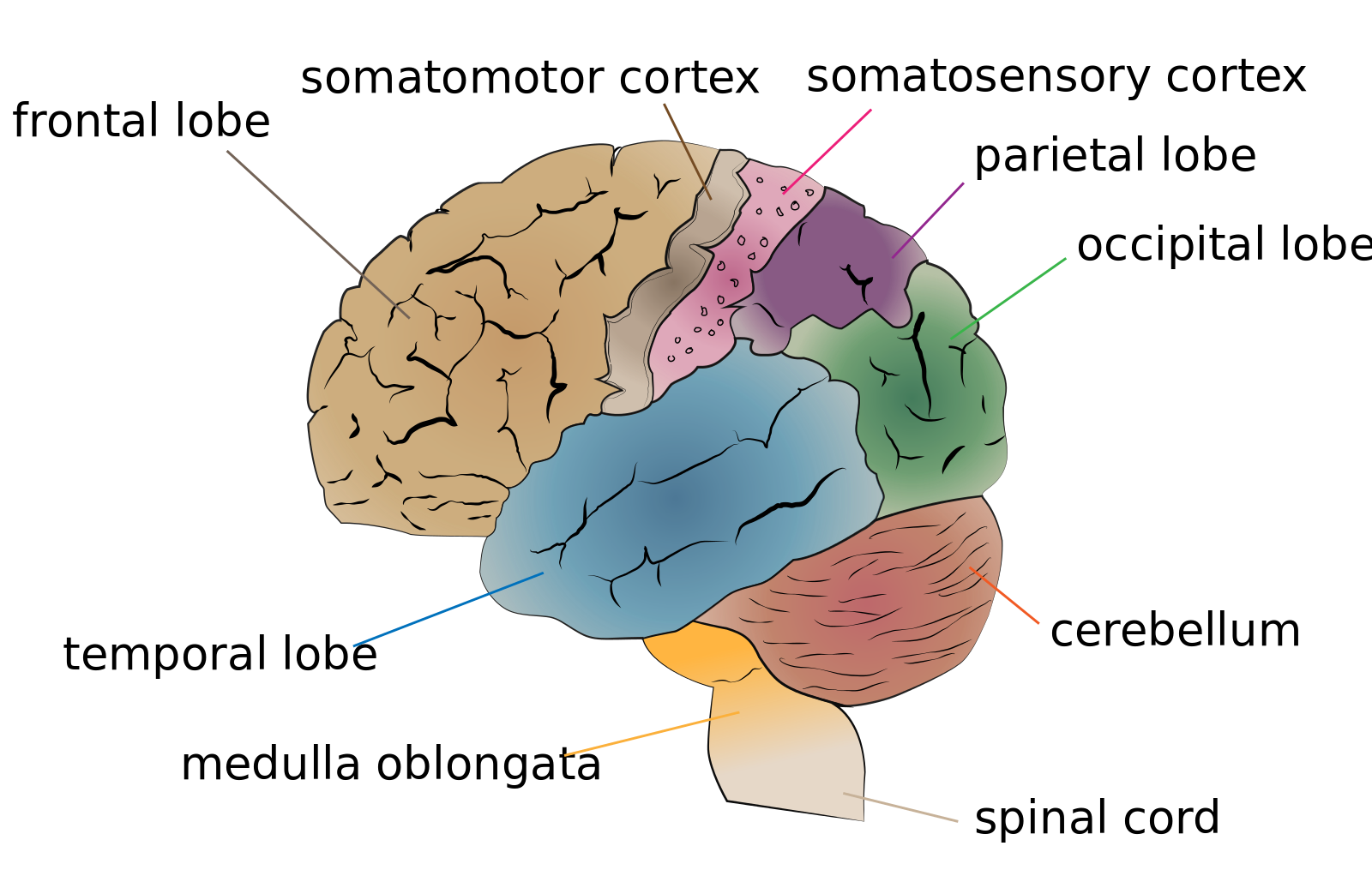
Diagram of the Brain
The brain gives us self-awareness and the ability to speak and move in the world. Its four major regions make this possible: The cerebrum, with its cerebral cortex, gives us conscious control of our actions. The diencephalon mediates sensations, manages emotions, and commands whole internal systems. The cerebellum adjusts body movements, speech.
Label the Brain
show/hide words to know What Are the Parts of the Brain? Every second of every day the brain is collecting and sending out signals from and to the parts of your body. It keeps everything working even when we are sleeping at night. Here you can take a quick tour of this amazing control center.
Brain Drawing With Labels at GetDrawings Free download
7,46,745 The Human Brain On average, an adult brain weighs between 1.0 kg - 1.5 kg. It is mainly composed of neurons - the fundamental unit of the brain and nervous system. Recent estimates have suggested that the brain contains anywhere between 86 billion to 100 billion neurons.
BRAIN DIAGRAM Unmasa Dalha
A well-labelled diagram of a human brain is given below for further reference. Structure And Function Of The Human Brain Parts Of The Human Brain The human brain is divided into three main parts: Forebrain. Midbrain. Hindbrain. These three main parts comprises many small parts. Forebrain The forebrain is also called as Prosencephalon.

Human Brain Diagram Labeled, Unlabled, and Blank
The basic structure of a neuron and an overall diagram of the human nervous system. Meninges : Coronal section A study of the meninges, ventricles, the circulation of cerebrospinal fluid and an illustration of the dura mater and falx cerebri. Ventricular system , Neuroanatomy : Lateral aspect
Brain diagram labeled
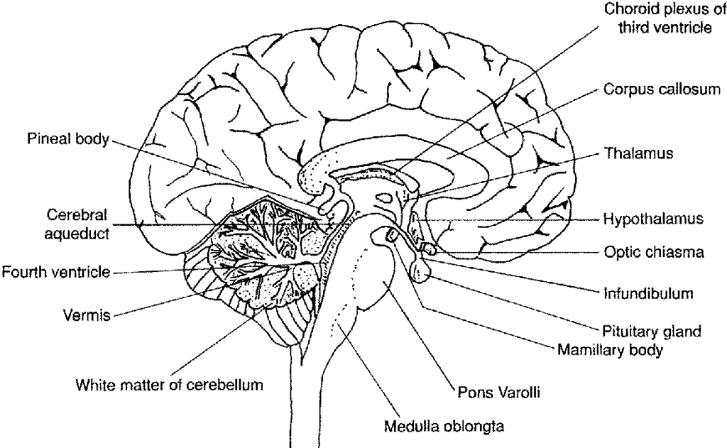
By: Tim Taylor Last Updated: Jul 30, 2020 2D Interactive NEW 3D Rotate and Zoom Anatomy Explorer HINDBRAIN AND MIDBRAIN Brain Stem Inferior Colliculus Medulla Oblongata Pons Quadrigeminal Lamina Superior Colliculus Cerebellum Cerebellar Peduncle 4th Ventricle Cerebral Aqueduct Choroid Plexus FOREBRAIN Diencephalon Choroid Plexus of 3rd Ventricle

Brain Model Labeled Brain anatomy, Anatomy and physiology, Brain models
Diagrams Diagrams are the perfect way to get orientated with a structure's detailed anatomy. Read on to see how we recommend using them. If you need some help with labeling the following diagrams, check out this video where we show you how to do it step-by-step: Labeled brain diagram