
Human Heart Diagram Unlabeled Tim's Printables
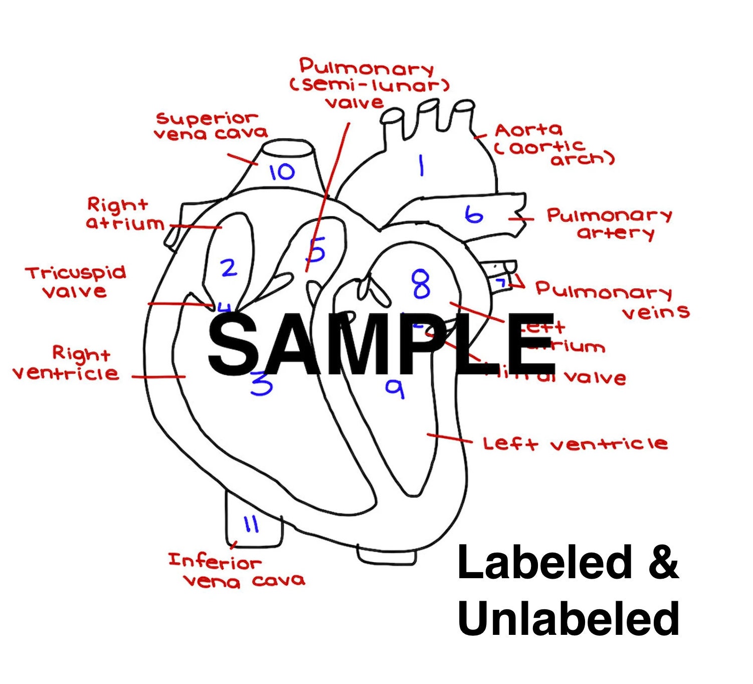
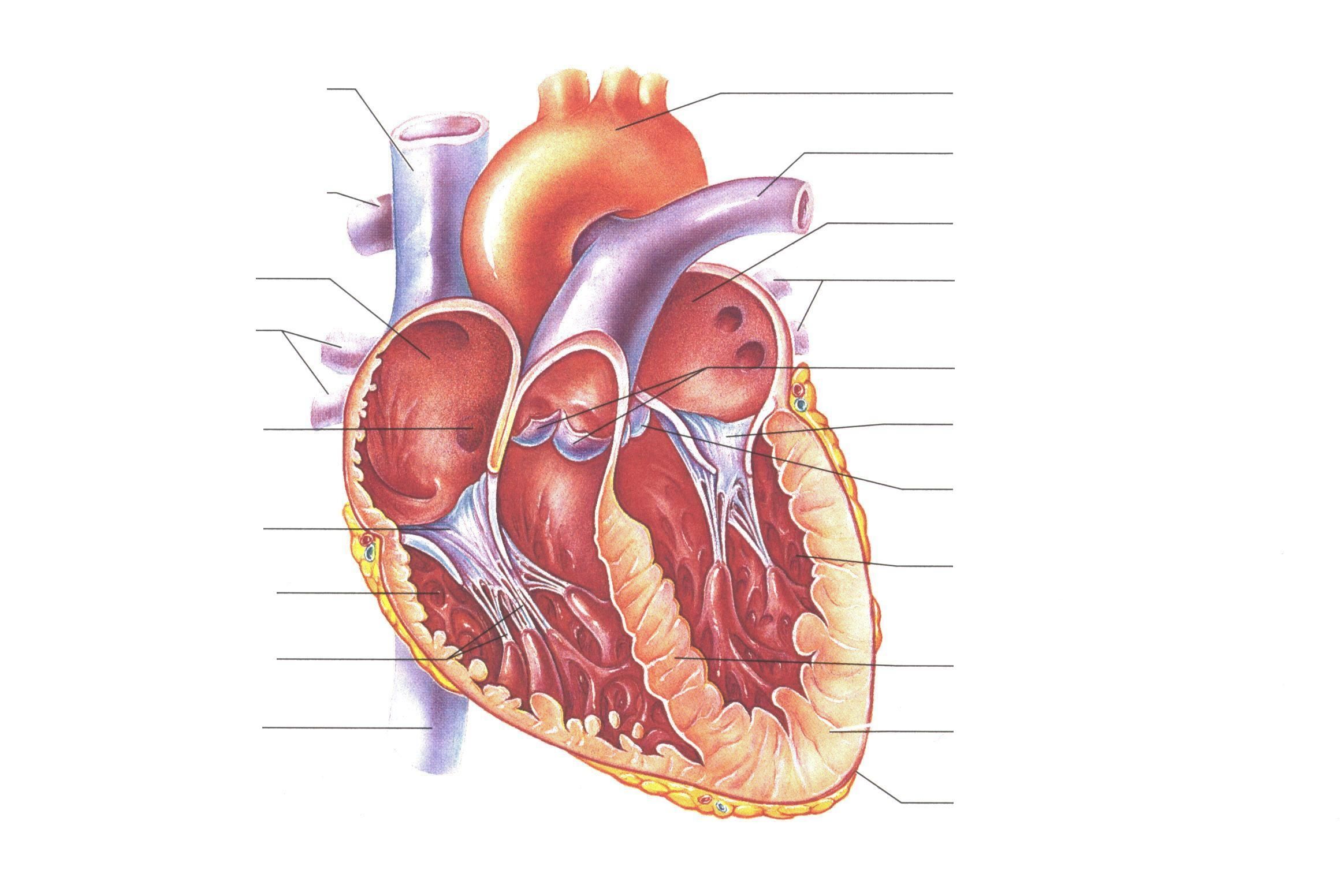
The human heart is primarily comprised of four chambers. The two upper chambers are called the atria, the remaining two lower chambers are the ventricles. The right and left sides of the heart are separated by a muscle called the "septum.". Both sides work together to efficiently circulate the blood.

The Heart Diagrams Labeled and Unlabeled 101 Diagrams
Selecting or hovering over a box will highlight each area in the diagram. For optimal viewing of this interactive, view at your screen's default zoom setting (100%) and with your browser window view maximised. See the Labelling the heart activity for additional support in using this interactive. Parts of the heart
The Heart Diagram Labeled and Unlabeled Worksheets Heart Etsy
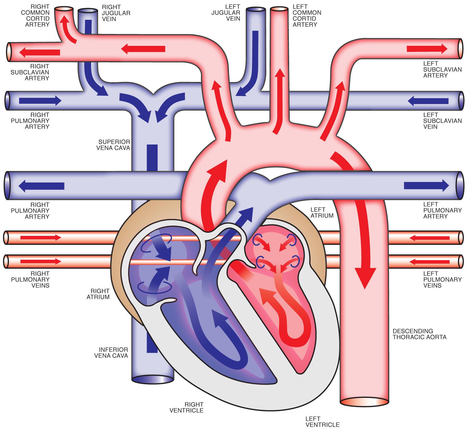
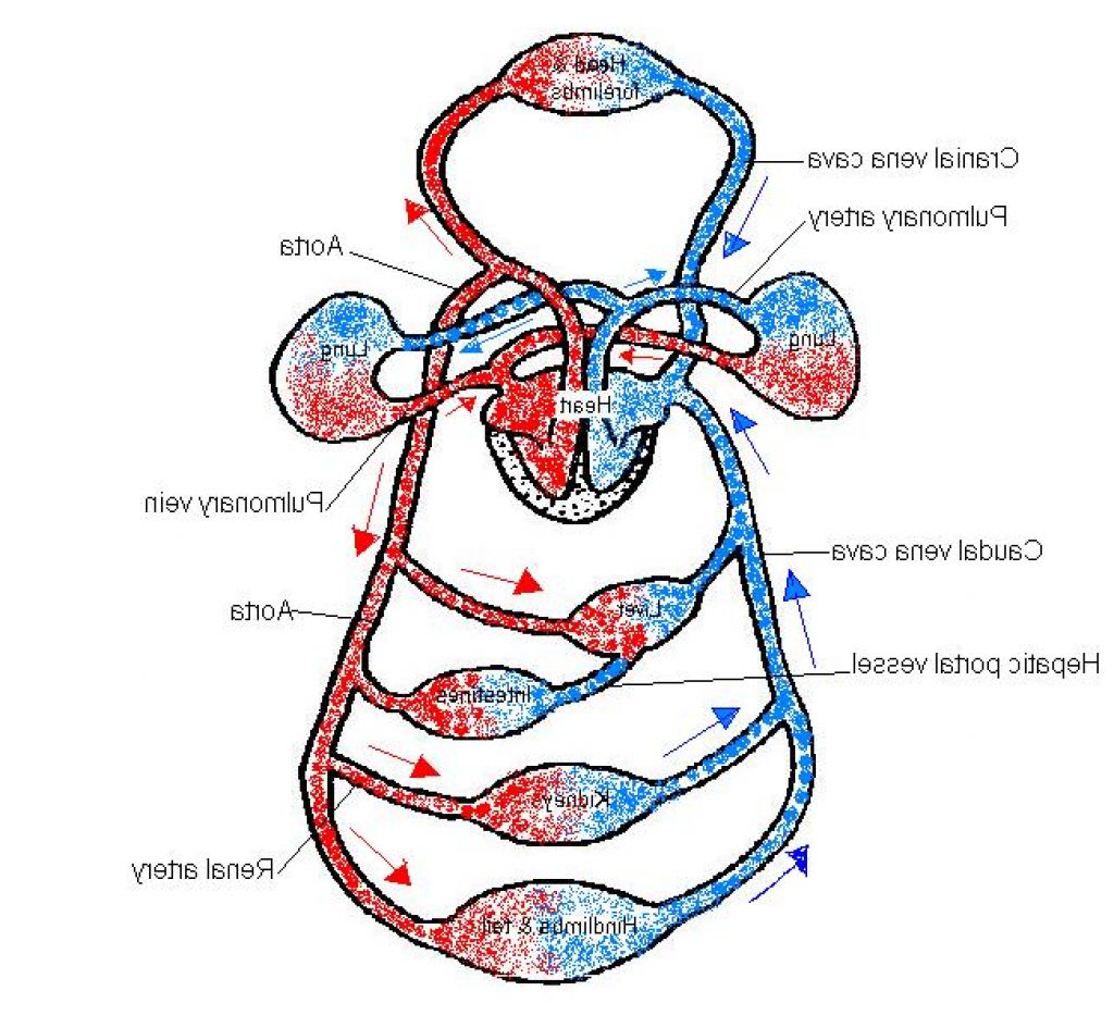
The cardiovascular system is a vital organ system which is quite literally at the centre of everything. Comprised of the heart, blood vessels and the blood itself, it is divided into two loops which both begin in the heart. The pulmonary circuit is responsible for exchanging blood between the heart and lungs for oxygenation, while the systemic circuit directs blood to the other tissues of the.
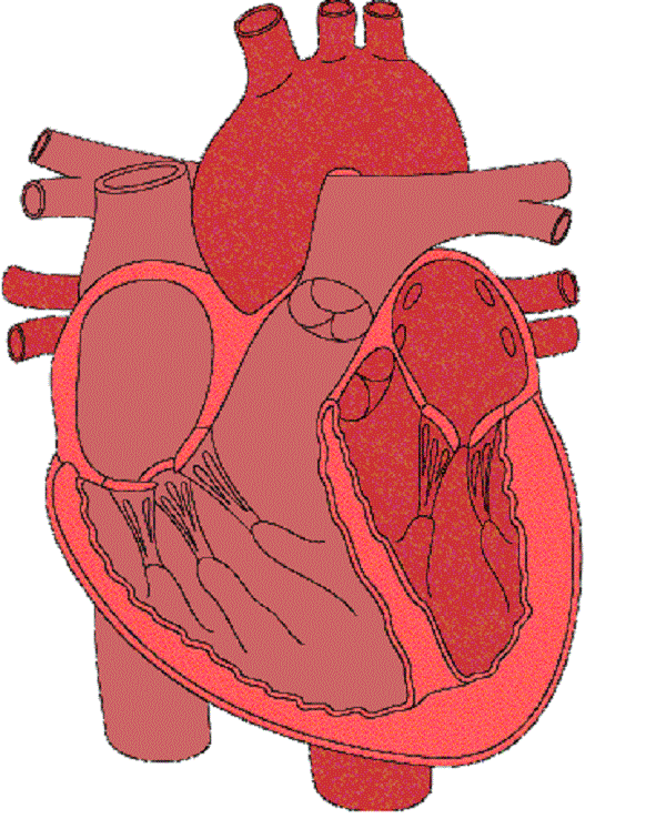
Unlabelled Diagram Of The Heart ClipArt Best
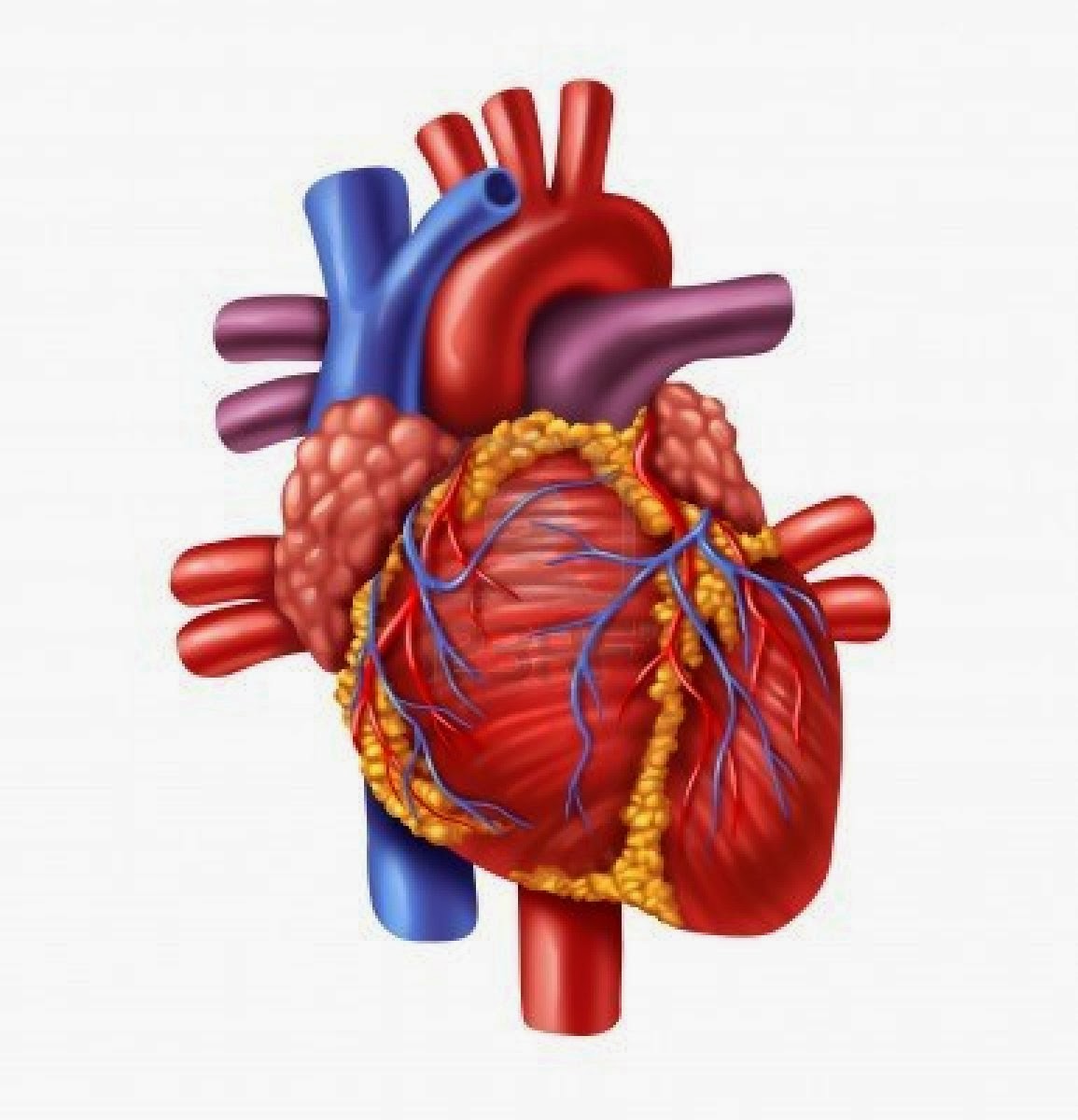
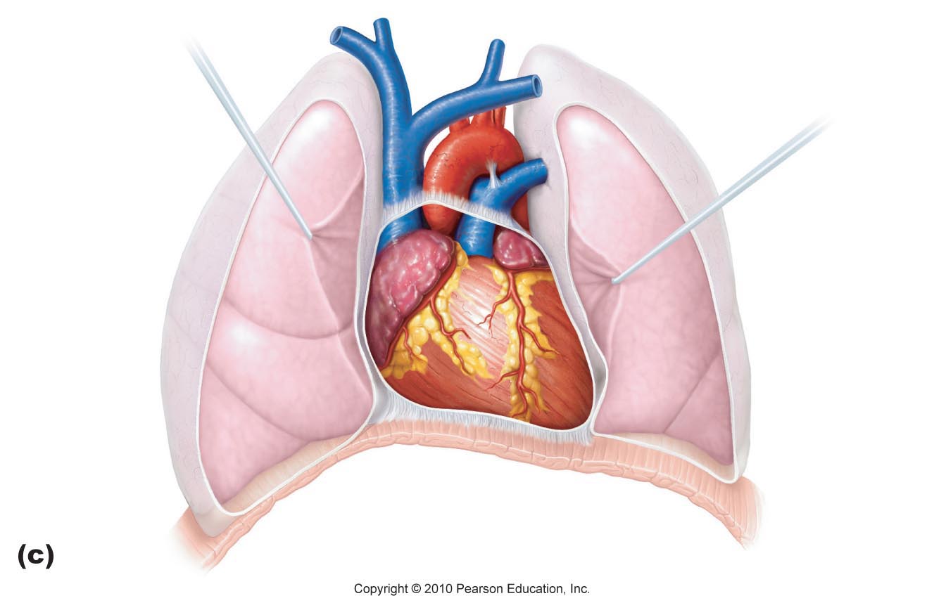
The heart is located in the thoracic cavity medial to the lungs and posterior to the sternum. On its superior end, the base of the heart is attached to the aorta,mycontentbreak pulmonary arteries and veins, and the vena cava. The inferior tip of the heart, known as the apex, rests just superior to the diaphragm.

Unlabeled Diagram Of The Heart General Wiring Diagram
The position of the heart in the torso between the vertebrae and sternum (see Figure 19.1.1 for the position of the heart within the thorax) allows for individuals to apply an emergency technique known as cardiopulmonary resuscitation (CPR) if the heart of a patient should stop. By applying pressure with the flat portion of one hand on the sternum in the area between the line at T4 and T9.
Heart Diagram Unlabeled Cliparts.co
heart, organ that serves as a pump to circulate the blood.It may be a straight tube, as in spiders and annelid worms, or a somewhat more elaborate structure with one or more receiving chambers (atria) and a main pumping chamber (ventricle), as in mollusks. In fishes the heart is a folded tube, with three or four enlarged areas that correspond to the chambers in the mammalian heart.
Unlabelled Diagram Of The Heart Cliparts.co
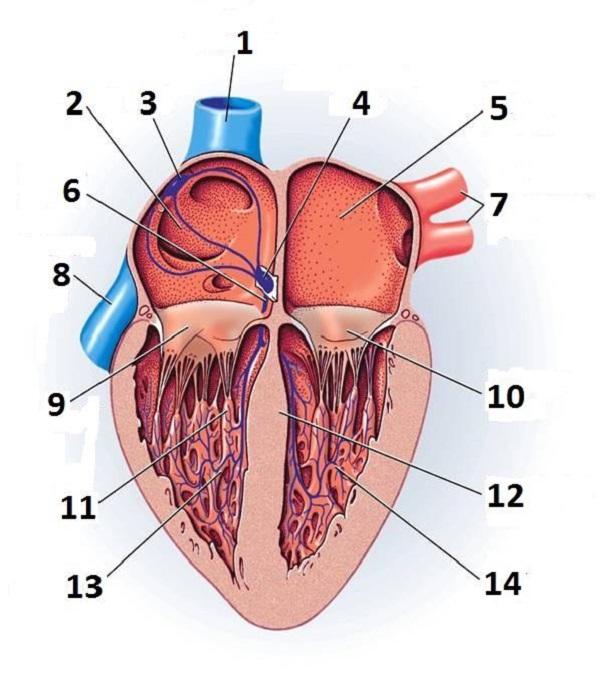
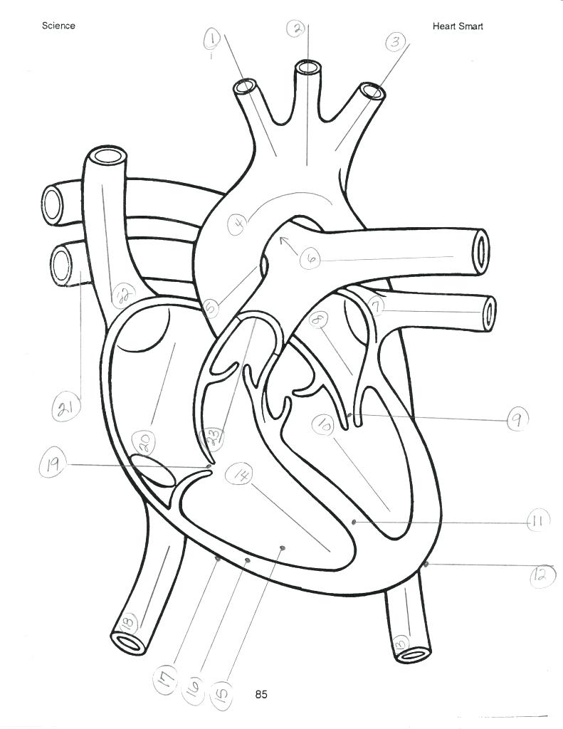
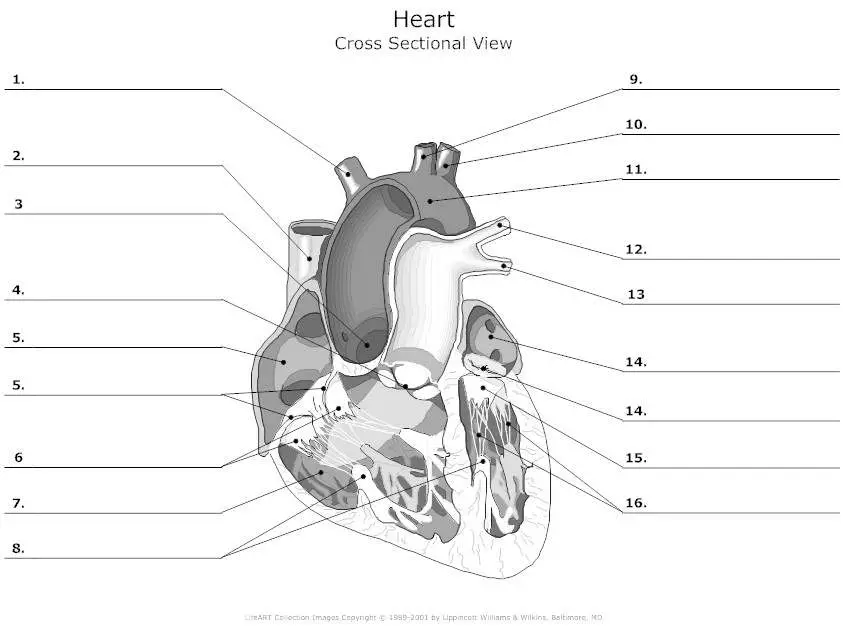
Don't forget to LABEL the parts of the heart on the diagram! 1. Compare the location of the tricuspid and bicuspid. 2. Compare the direction of blood flow in the pulmonary artery to the pulmonary vein. 3. Mitral regurgitation is a heart condition that occurs when the mitral valve does not close fully. Based on your knowledge of the heart.

Module 3 Cardiovascular Assessment and Health Promotion at Mount Royal
English: Diagram of the human heart, without identifying labels. Date: 29 July 2015: Source: Own work, based on Image:Diagram of the human heart (cropped).svg: Author: Pereru: Licensing [edit] I, the copyright holder of this work, hereby publish it under the following license:
The best free Diagram drawing images. Download from 3558 free drawings
Function and anatomy of the heart made easy using labeled diagrams of cardiac structures and blood flow through the atria, ventricles, valves, aorta, pulmonary arteries veins, superior inferior vena cava, and chambers. Includes an exercise, review worksheet, quiz, and model drawing of an anterior view (frontal section) of the heart in order to.
Unlabelled Diagram Of The Heart ClipArt Best
A heart diagram is a visual representation of the different parts of the heart, including the chambers, valves, and major blood vessels. Why is it important to understand your heart diagram? Understanding your heart diagram can help you better understand how your cardiovascular system works and what you can do to keep it healthy.
Heart Diagram Unlabeled Cliparts.co
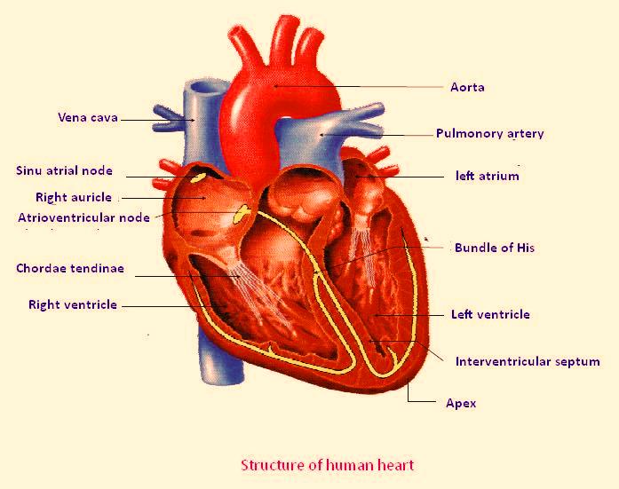
The heart has three layers. They are the: Epicardium: This thin membrane is the outer-most layer of the heart. Myocardium: This thick layer is the muscle that contracts to pump and propel blood.
Heart Diagram Unlabeled Cliparts.co
Diagram of Heart. The human heart is the most crucial organ of the human body. It pumps blood from the heart to different parts of the body and back to the heart. The most common heart attack symptoms or warning signs are chest pain, breathlessness, nausea, sweating etc. The diagram of heart is beneficial for Class 10 and 12 and is frequently.

Human Heart Unlabeled ClipArt Best
The diagram is clear and simple and contains large text. Furthermore, a blank human heart diagram has been provided, allowing you to fill in the blanks for an incoming test or quiz. These human heart diagrams are available free of charge as medium resolution jpegs. Simple click the image links below to go to the Printables Libary download page.
Heart Diagram Unlabeled Cliparts.co
Heart Review Quiz: Once the activity is completed and cleaned up, show students an unlabeled diagram of the heart. Do this as a handout or projected overhead transparency or PowerPoint® slide. If the blank diagram is shown to the entire class at once, point out the various parts of the heart, including the following structures: left and right.
Heart Diagram Unlabeled Cliparts.co
Heart anatomy. The heart has five surfaces: base (posterior), diaphragmatic (inferior), sternocostal (anterior), and left and right pulmonary surfaces. It also has several margins: right, left, superior, and inferior: The right margin is the small section of the right atrium that extends between the superior and inferior vena cava .
Unlabelled heart diagram
1. To find a good diagram, go to Google Images, and type in "The Internal Structure of the Human Heart". Find an image that displays the entire heart, and click on it to enlarge it. [1] 2. Find a piece of paper and something to draw with. Start with the pulmonary veins.